RESPIRATORY FAILURE
- Notes about respiratory monitoring
- O2 goal may be lower for chronic retainer
- Beware of CO, methemoglobin, abnormal hemoglobin, which may falsely elevate O2 measurement
- Pulse Ox is less reliable at values below 80
- Nail polish, dyes, (methylene blue), tremor may cause artifact affecting pulse ox measurement; look for good waveform
- Rough VBG-to-ABG conversion
- Subtract 8 from PaCO2
- Add 0.04 to pH
- Rough SaO2 to PaO2 conversion:
| SaO2 (%) | 80 | 84 | 88 | 92 | 96 | 100 |
| PaO2 (mmHg) | 44 | 49 | 55 | 65 | 86 | 145 |
- Consider intubation when:
- Respiratory failure (hypoxemic and hypercapneic)
- Inability to protect airway (GCS < 8)
HYPOXEMIC RESPIRATORY FAILURE
A failure of OXYGENATION
- Notes: alveoli are more compliant at lung bases, and V and Q are higher at bases
- Hypoxemia symptoms: for previously healthy patient:
- PaO2 < 50mm Hg: malaise, light-headedness, nausea, vertigo, incoordination, confusion
- PaO2 < 35mm Hg: decreased renal blood flow, decreased UOP, bradycardia, conduction blocks, lactic acidosis
- PaO2 < 25mm Hg: LOC, respiratory depression
- Hyperoxia symptoms:
- O2 replacing Nitrogen à atelectasis, poor lung compliance, ROS
HYPERCAPNEIC RESPIRATORY FAILURE
A failure of VENTILATION
- Mechanisms: airflow obstruction, muscular weakness/ineffective musculature, inadequate ventilatory drive, increased ventilation requirement
- Note: can follow minute ventilation (f x VT in liters) to trend PaCO2. If trending ABG’s, goal should be normal pH, not normal PaCO2 or PaO2
- Hypercapnea symptoms:
- Cerebral vasodilation à increased ICP
- Adrenergic stimulation à increased CO, PVR
- Decreased tissue metabolism
- Improved surfactant function
- Prevents protein nitration
- Oxygen can worsen hypercapnia by:
- Increased V/Q mismatch
- Suppressed central hypoxemic drive
- Haldane effect:
- In RBC’s: CO2 + H2O ó H2CO3 ó H+ + HCO3–.
- Oxygenation of Hb promotes dissociated of H+ from Hb, which shifts the bicarbonate buffer equilibrium toward CO2 formation, so CO2 is released from RBC’s.
- Therefore, high plasma [O2] causes Hb to release CO2, and diseased lungs may not be able to adequately increase alveolar ventilation to breathe off this CO2.
Supplemental Oxygen
Escalation of oxygen therapy
| Device | Flow rate (L/min) | FiO2 (%) approx. | PEEP? |
| Nasal cannula | 1 | 24 | No |
| 2 | 28 | ||
| 3 | 32 | ||
| 4 | 36 | ||
| 5 | 40 | ||
| Face mask | 5 | 40 | No |
| 6-7 | 50 | ||
| 7-8 | 60 | ||
| Venturi mask | 3-15 | 24 – 50 | No |
| Non-rebreater | 12 – 15 | > 90 | No |
| High flow nasal cannula | Yes | ||
| CPAP | Yes | ||
| Endotracheal tube | 21 – 100 | Yes |
Non-invasive positive pressure ventilation (NIPPV)
- CPAP: continuous positive airway pressure
- CHF (creates pressure gradient from chest to periphery, allowing for decreased afterload, does not push fluid out of lungs)
- BPAP: bi-level positive airway pressure (IPAP/EPAP)
- COPD, pressure difference allows for ventilation (think as external diaphragm)
- Contraindications to NIPPV:
- Inability to protect airway
- AMS (GCS </= 8), poor compliance, seizure, stroke
- Fluid that could be forced into airway: vomiting, epistaxis, upper GI bleed, secretions (avoid in pneumonia unless patient is immunocompromised)
- Severe facial trauma or burn
- Pneumothorax, penetrating chest trauma
- CSF leak
Respiratory Glossary
- Positive end-expiratory pressure (PEEP): pressure applied to alveoli at end-expiration
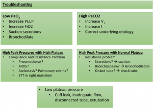
- Plateau pressure (Pplat): pressure applied to small airways/alveoli at end-inspiration during a period of no airflow (inspiratory hold). Reflects lung compliance.
- Note: Pplat more accurately measured when flow rate is constant, so less reliable in PRVC—if measuring in PRVC, check multiple times and average
- Peak pressure (Ppeak): the highest pressure needed to move a volume of gas through major airways into the lung. Reflects airway resistance (but is a combination of compliance and resistance).
- Airway resistance is proportional to viscosity of inspired gas and length of airway, and inversely proportional to radius of airway (Poiseuile’s law)
- Airway resistance = (pressure in mouth – pressure in alveoli)/airflow (Ohm’s law)
- Mean airway pressure: average airway (large and small) pressure throughout ventilatory cycle
- Inspiratory to expiratory (I:E) Ratio: ratio of inspiratory to expiratory time in the ventilatory cycle
- Lung compliance (C) = tidal volume DV / transpulmonary pressure DP
Mechanical Ventilation
Peri-intubation check-list:
- Induction agent, Consideration of anxiolytic and analgesic, Paralytic (MUST HAVE ATTENDING PRESENT)
- pre and peri-intubation oxygenation with high-flow NC in hypoxemic patients
- Calculate ideal body weight to determine VT; lung protective strategy preferred even in non-ARDS patients (VT < 8 mL/kg IBW)
- Check for bilateral breath sounds, chest rise, end-tidal CO2, vitals immediately post-intubation
- Order stat CXR to check position of tube (should be 4cm above carina)
- No need for daily CXR in intubated patients; on-demand strategy associated with fewer CXR’s and no change in patient outcomes: ventilator days, length of stay, or mortality5
- Order ABG 30 minutes post-intubation (you do not need to use PO2 to wean FiO2, simply use pulse ox)
Modes:
- Variables
- Control: pressure (P), volume (TV), or both
- Trigger: elapsed time, decrease in intrathoracic pressure, negative inspiratory flow (either time-triggered or patient-triggered)
- Cycle: respiratory rate (f), inspiratory to expiratory (I:E) ratio
- FiO2 (0.21 – 1.0)
- PEEP
| Mode | Ventilator controls… | Patient controls… | Notes: |
| (AC) Volume Control | VT, f, PEEP, FiO2, | f above vent | Airway pressures vary depending on lung and chest wall compliance, airway resistance |
| (AC) Pressure Control | Pinsp, I:E, f, PEEP, FiO2, | f above vent | VT varies depending on lung compliance, airway resistance |
| Set MV cannot be guaranteed | |||
| PRVC | VT, Ppeak, f, FiO2, PEEP | f above vent | Delivered VT will be lower than set VT if Ppeak exceeded, and may be higher than set VT. |
| (pressure-regulated volume control) | |||
| Pressure Support | Pinsp FiO2, PEEP | VT, f, flow rate | Used for spontaneous breathing trials (SBT’s) |
| SIMV (synchronous intermittent mechanical ventilation) | VT of vent-triggered breaths, f, PEEP, FiO2 | VT of extra breaths, f above vent | Used commonly in surgical ICU’s |
| APRV | Phigh, Plow, Thigh, Tlow, FiO2 | Spontaneous breathing allowed throughout cycle | Intentional auto-PEEP to avoid derecruitment |
| (airway pressure release ventilation) | Allows alveolar recruitment while minimizing ventilator-induced lung injury. May be useful in ARDS6 |
Weaning
- Reason for intubation improved
- Hemodynamically stable
- FiO2 </= 40%, PEEP </= 5
- Good cough and gag
- Discontinue sedation, hold tube feeds in advance (or suction out)
- SBT (spontaneous breathing trial): PS 10/5, 5/5, 0/5, or t-piece trial
- Daily spontaneous awakening trial (SAT) + spontaneous breathing trial (SBT) improve outcomes: more ventilator-free days, earlier discharge, decreased 1-year mortality
- 30 min – 2 hours sufficient
- RSBI (rapid shallow breathing index) = f/VT
- RSBI >/= 105 portends likelihood of unsuccessful extubation
- NIF (negative inspiratory force) less than negative 20
- FVC >/= 10 mL/kg IBW
- Cuff leak: deflate ETT cuff; VT should decrease by 20% (because volume is being lost around the tube. No cuff leak indicates laryngeal edema
- Consider extubating to BiPAP
- Once extubated
- Swallow study (bedside is fine unless they fail that)
- If stridor, give 10mg dexamethasone IV STAT, reintubate earlier rather than later

